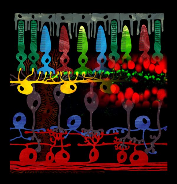
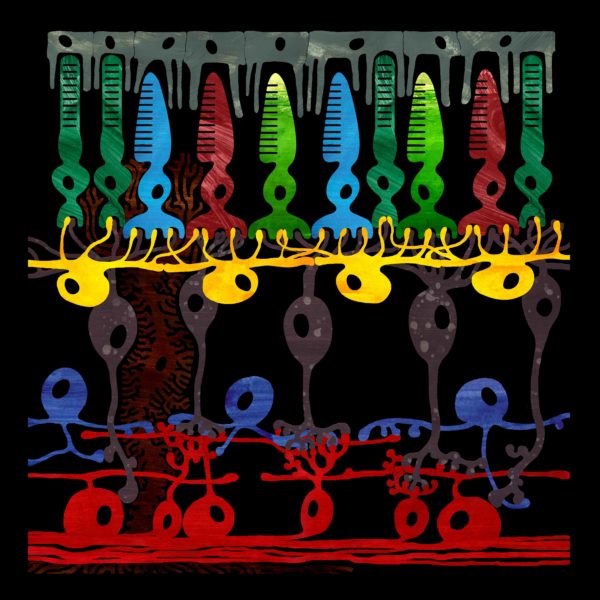
Schematic drawing of the vertebrate retina combined with the immunolocalization of the ecto-ATPase NDTPdase1 (green). The top grey cells are pigment epithelial cells. Right underneath are the cone photoreceptors depicted in red, green and blue. The dark green cells are rod photoreceptors. The yellow cells are horizontal cells and thedark purple cells are bipolar cells. Amacrine cells are depicted in blue-purple and the red cells at the bottom are the ganglion cells. The dark brown cell in the background is a Müller cell.
A more detailed explanation of the schematic drawing and the different cell types can be found on the education images page.
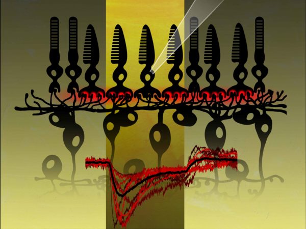
The brain never ‘sees’ a raw image of the visual world. The outside world contains a lot of different shapes, colours, textures, light levels and movement. Clearly there is a lot of information for the brain to deal with and the retina is therefore doing a good bit of prework before sending signals to the brain. Substantial processing of the incoming visual information is already taking place at the first retinal synapse. Here, horizontal cells (yellow) modulate the signal from photoreceptors to bipolar cells via a negative feedback pathway. The process underlying this inhibitory interaction is an unexpected combination of an extremely fast and a slow mechanism. The slow component is a newly discovered form of synaptic communication involving pannexin 1 channels and extracellular ATP hydrolysis by ecto-ATPases (green label, image-right). The mechanism inhibits by changing the pH buffer capacity in the synaptic cleft.
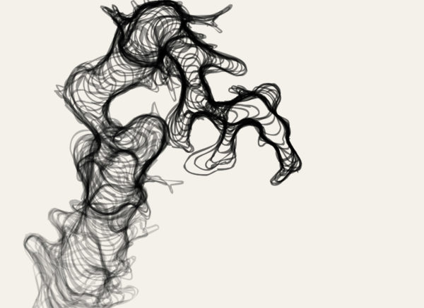
This image is is based on the movements of neutrophils, the most common type of white blood cell. Neutrophils respond to certain chemicals and migrate towards the highest concentration, a mechanism called chemotaxis. Bacteria use this mechanism to move themselves towards valuable nutrients to feed on and sperm cells use it to find their way towards the egg for fertilisation. Neutrophils are part of the immune system and use chemotaxis to guide themselves to a site of inflammation. Once there, they are attracted to bacteria and will chase them around until they are close enough to absorb and kill them. In the animation the neutrophil is first attracted to a virtual site of inflammation and subsequently starts chasing an imaginary bacteria. However, I have left everything out except from the neutrophil itself. The animation is then collapsed to show the whole path at once.
This website uses cookies to improve your experience. If you continue to use this website we will assume that you are happy with this. OKTerms & conditions
Privacy & Cookies Policy
Privacy Overview
This website uses cookies to improve your experience while you navigate through the website. Out of these cookies, the cookies that are categorized as necessary are stored on your browser as they are essential for the working of basic functionalities of the website. We also use third-party cookies that help us analyze and understand how you use this website. These cookies will be stored in your browser only with your consent. You also have the option to opt-out of these cookies. But opting out of some of these cookies may have an effect on your browsing experience.
Necessary cookies are absolutely essential for the website to function properly. This category only includes cookies that ensures basic functionalities and security features of the website. These cookies do not store any personal information.
Any cookies that may not be particularly necessary for the website to function and is used specifically to collect user personal data via analytics, ads, other embedded contents are termed as non-necessary cookies. It is mandatory to procure user consent prior to running these cookies on your website.