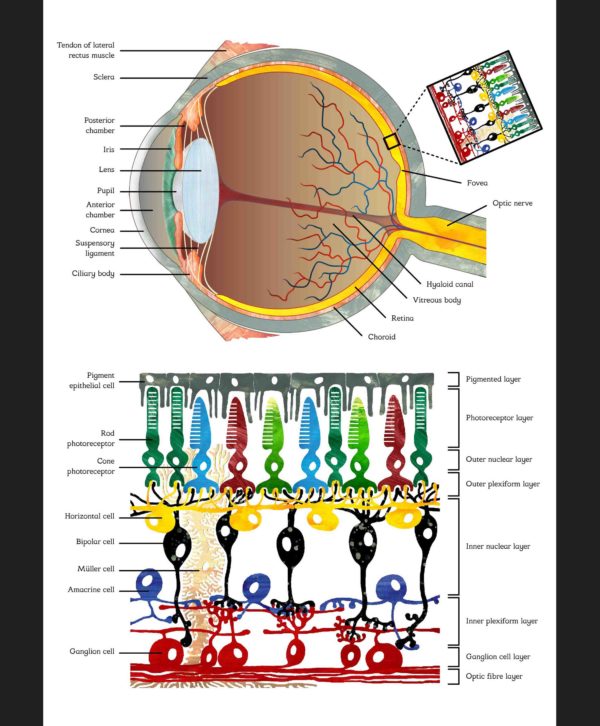
When light enters the eye, a visual image is focussed on a structure at the back of the eye that we call the retina. This structure contains several layers of neurons and is connected to the brain via a bundle of fibres we call the optic nerve. However, the brain does not simply receive the raw retinal image. The incoming visual information is first heavily processed by the various layers of the retina. The cells that first detect the light are called photoreceptors. There are two types: cones, which are highly concentrated in the fovea for detail and mainly function during the day, and rods that primarily function in very dark conditions because they are extremely sensitive and can even respond to a single photon. In the schematic rods are depicted as dark green. They have no role in colour vision which explains why it is so hard to make out colours in low light conditions such as during the night. Cones come in three types in humans, each one is most sensitive to a different colour: red, blue and green. In the schematic the different types of cones are represented by their corresponding colours.
After the light is picked up by the photoreceptors the image is further processed by several other neuronal cell types. Incredibly, substantial processing is already taking place at the point where the cones communicate with two cell types in the next layer, which are known as horizontal cells (yellow) and bipolar cells (black). The point at which two cells communicate is called a synapse and so we refer to this as the first retinal synapse. The mechanisms underlying the processing at the first retinal synapse formed the topic of my PhD study.
The signals from bipolar cells are received by the ganglion cells (red) that form the last layer in the retina and do the last bit of processing, turning the signals into a message that the brain can understand and further process. However, we should not skip over the amacrine cells (purple) that are, like the horizontal cells, not part of the direct path from photoreceptor to optic fibre, but modulate and integrate the signals in this path in various ways, and connect to both bipolar cells and ganglion cells. There are many different types of amacrine cells that all have important roles in interpreting the raw image.
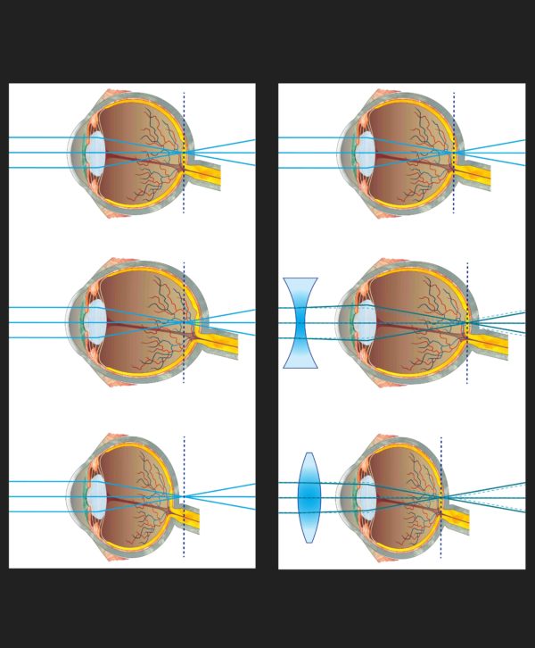
Schematic image of normal eyesight, myopia (nearsightedness) and hyperopia (farsightedness) and their correction using concave and convex lenses respectively
Light entering the eye is projected by the cornea and lens on a layered neuronal network that covers the inner back wall of the eye, the retina (see above). The schematic on the left explains how a slight elongation or shortening of the eyeball prevents the projection to be properly focussed on the retina. The schematic on the right shows how this can be corrected for using glasses with lenses that are shaped to slightly change the light-path before passing the lens of the eye.
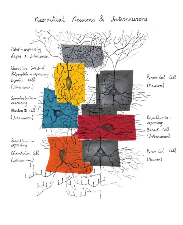
Drawing of neocortical neurons and several sub-types of interneurons
Originally this image did not contain text as it was meant as an artist impression of a micro-network in the neocortex. A detailed explanation for this painting can therefore be found on the page about neuron drawings. However, because the design was based on currently used classifications of neuronal types in neuroscience, I made a version with text to give people an idea about the different neuron types and what they look like.
This website uses cookies to improve your experience. If you continue to use this website we will assume that you are happy with this. OKTerms & conditions
Privacy & Cookies Policy
Privacy Overview
This website uses cookies to improve your experience while you navigate through the website. Out of these cookies, the cookies that are categorized as necessary are stored on your browser as they are essential for the working of basic functionalities of the website. We also use third-party cookies that help us analyze and understand how you use this website. These cookies will be stored in your browser only with your consent. You also have the option to opt-out of these cookies. But opting out of some of these cookies may have an effect on your browsing experience.
Necessary cookies are absolutely essential for the website to function properly. This category only includes cookies that ensures basic functionalities and security features of the website. These cookies do not store any personal information.
Any cookies that may not be particularly necessary for the website to function and is used specifically to collect user personal data via analytics, ads, other embedded contents are termed as non-necessary cookies. It is mandatory to procure user consent prior to running these cookies on your website.